Abstract
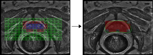
Segmentation of the prostate boundary on clinical images is useful in a large number of applications including calculation of prostate volume pre- and post-treatment, to detect extra-capsular spread, and for creating patient-specific anatomical models. Manual segmentation of the prostate boundary is, however, time consuming and subject to inter- and intra-reader variability. T2-weighted (T2-w) magnetic resonance (MR) structural imaging (MRI) and MR spectroscopy (MRS) have recently emerged as promising modalities for detection of prostate cancer in vivo. MRS data consists of spectral signals measuring relative metabolic concentrations, and the metavoxels near the prostate have distinct spectral signals from metavoxels outside the prostate. Active Shape Models (ASM’s) have become very popular segmentation methods for biomedical imagery. However, ASMs require careful initialization and are extremely sensitive to model initialization. The primary contribution of this paper is a scheme to automatically initialize an ASM for prostate segmentation on endorectal in vivo multi-protocol MRI via automated identification of MR spectra that lie within the prostate. A replicated clustering scheme is employed to distinguish prostatic from extra-prostatic MR spectra in the midgland. The spatial locations of the prostate spectra so identified are used as the initial ROI for a 2D ASM. The midgland initializations are used to define a ROI that is then scaled in 3D to cover the base and apex of the prostate. A multi-feature ASM employing statistical texture features is then used to drive the edge detection instead of just image intensity information alone. Quantitative comparison with another recent ASM initialization method by Cosio showed that our scheme resulted in a superior average segmentation performance on a total of 388 2D MRI sections obtained from 32 3D endorectal in vivo patient studies. Initialization of a 2D ASM via our MRS-based clustering scheme resulted in an average overlap accuracy (true positive ratio) of 0.60, while the scheme of Cosio yielded a corresponding average accuracy of 0.56 over 388 2D MR image sections. During an ASM segmentation, using no initialization resulted in an overlap of 0.53, using the Cosio based methodology resulted in an overlap of 0.60, and using the MRS-based methodology resulted in an overlap of 0.67, with a paired Student’s t-test indicating statistical significance to a high degree for all results. We also show that the final ASM segmentation result is highly correlated (as high as0.90) to the initialization scheme.
Link for the publication: https://www.sciencedirect.com/science/article/abs/pii/S136184151000112X

