
Abstract
The use of mobile devices is revolutionizing the way we communicate, interact, are entertained, and organize our lives. With healthcare in general and radiology in particular becoming increasingly digital, the use of such devices in radiologic practice is inevitable. This article reviews the current status of the use of mobile devices in the clinical practice of radiology, namely in emergency teleradiology. Technical parameters such as luminance and resolution are discussed. The article also discusses the benefits of such mobility vis-à-vis the current limitations of the technologies available.
Introduction
The practise of radiology is slowly but surely evolving toward the universal adoption of teleradiology by radiologists and radiology groups across the world, as can be seen from the literature published on the subject.[1,2] Logically speaking, this would make perfect sense. When image data is digital, similar to all other digital information, then there seems no reason why should it not be transmitted across a network to facilitate timeliness of reporting that is in the interests of improved patient care. Of course there are complex legal, regulatory issues, etc., that must be addressed, but in essence, from a technology perspective, radiology is today a specialty that it has been shown can be practised safely and accurately from off-site.[3]
iPad
The most recent change in the dynamic has been the introduction of portable handheld devices, such as tablet PCs and smartphones, which allow for on-the-go interpretation of medical images, as opposed to the use of an office-based workstation.[4,5] At the forefront of this mobile revolution have been the legacies of Steve Jobs, the iPad and iPhone.
Since the inception of the iPad in 2010, it has been a game changer in the way people access the information, work, play, communicate on the move. There have been several other tablets based on the Android system and there is now the Playbook from Blackberry. However, the iPad is clearly the leading force in the tablet market.[6] The picture archiving and communication system (PACS) applications that currently run on mobile handheld devices include Merge Mobile, OsiriX, iClarity, Mobile MIM, ResolutionMD, Fujifilm Synapse, and RadSpa.
With web-based access to quality images now becoming a reality and the increasing penetration of wireless broadband of 2G and 3G, mobile radiology systems have the potential to dramatically improve the quality of patient care by facilitating instant diagnosis. The availability of high-speed bandwidth on iPads and mobile tablet devices allows for immediate transfer of images of critical value in the emergency setting.
A secondary arguable benefit of mobile tablet based radiology systems is their potential to increase radiologists’ productivity by allowing them to work while on-the-go, as while traveling, thereby reducing their downtime and helping ease the radiologist shortage.
As radiologists, however, it is important for us to understand and be aware of the benefits as well as potential pitfalls of the use of such technologies before embracing their use. There are some unique benefits that iPad and other mobile tablets bring to the radiology practice, which we believe will increase the adoption of these tablets in medical imaging.
Speed of Image Download
The availability of high-speed wireless bandwidth such as 2G and now 3G on mobile tablet devices allows for immediate transfer of images to tablets enabling “on the move” teleradiology, which is of great benefit in the emergency setting, as in the evaluation of acute stroke or acute abdominal emergencies.
Image Resolution and Display
The key question that arises is whether the images displayed on an iPad or other mobile tablets are of diagnostic quality.
Using a tablet PC, the radiologist is able to read the scans in high resolution with very little panning. The web-based application allows the reader to zoom and adjust the window level, contrast, and brightness of the image. The radiologist will be evaluating actual raw DICOM image data and not just JPEG snapshots. Depending on the application, size, and density measurement tools, and annotation features may also be available, allowing for a complete evaluation.
FDA has approved one application on the iPad for diagnostic viewing of medical images (with the obvious exception of mammography).[7]
The iPad’s 9.7-inch display provides 1024 × 768-pixel resolution at 132 pixels per inch (PPI). The larger display allows images to be displayed at or closer to their native resolution.
The iPad has a maximum luminance of 270 cd/m2, which, while being higher than the average of 150-200 cd/m2 seen in commercially available displays, is much lower than the average primary interpretation workstation display that has an average maximum luminance of 500-600 cd/m2. The iPad’s minimum luminance is 0.3 cd/m2, which yields a contrast ratio of 900:1 for a portable device. In that sense, the display of medical images on an iPad is superior to that of an off-the-shelf workstation displays but below that of the medical grade monitors.[8]
Portability and Mobility
The iPad with its sleek design and convenient form factor can be easily carried in a briefcase and used by radiologists to view medical images at the point of care without being tied to their workstations. A kerbside discussion between a radiologist and a surgeon can be greatly enhanced if the radiologist is able to pull out his or her iPad from the briefcase and demonstrate the actual imaging findings. Similarly, the patient experience is improved when the physician can review the results of the scan with the patient at his or her bedside with the images at hand.
“Always On” Feature
The iPad turns on easily and instantly unlike desktops or even laptops which need significant boot up time to be able to open up and review the medical images. In the setting of an acute emergency such as stroke or tension pneumothorax, every minute is critical and this can be an important benefit.
Efficiency Gain
While for radiologists, the ability to have immediate and mobile access to images is obviously of value, for clinicians as well, having portable access to secure and complete patient information in the hospital information system (HIS) along with the medical images will lead to considerable efficiency gains.[9]
Other Issues/Questions
Are tablets secure?
The health insurance portability and accountability Act (HIPAA) requires that the images are transferred securely and are accessed by authenticated users. The iPad and tablet applications for radiology need to ensure they use the secure socket layer (SSL) protocol or virtual private network (VPN) for transferring the medical images and the access to these images is login controlled. The images are not actually stored on the iPad, but are always accessed from the server through the login-based interface. Login timeout restricts the amount of time that the images are physically displayed on the device’s monitor. Ultimately, it is of course the responsibility of the radiologist or physician to ensure the physical security of his/her portable device.
Workflow issues
While, as we have seen, the iPad provides the adequate display resolution and clarity to interpret medical images, how does its actual use fit in to the clinical workflows of radiologists and physicians? Can one actually report an entire worklist of scans using an iPad?
Applications are currently available which provides a continuation of the regular workstation workflow with the iPads and tablets. The worklist on the iPad is in real-time synchronization with the worklist on the desktop, and hence identical in terms of user experience.
Additionally, the iPad application has the capability (using voice recognition tools) to either have the dictated reports instantly transcribed on the iPad itself or else have the voice file recorded to be transcribed online by a remote medical transcriptionist.
The iPad application can be customized to display as much of the medical record of the patient as may be required.
Furthermore, iPad-based applications can work as a communication tool between the radiologist, the referring physician, and even the patient.
As a teaching tool
Web-based teaching is greatly facilitated by the use of technologies such as tablet PCs. Saving of images into powerpoint presentations is facile with the iPad. In combination with the use of internet-based videoconferencing technologies, the iPad can greatly extend the reach of radiology training beyond the traditional classroom model. Of course, access to teaching websites is also facilitated by the use of portable tablet devices.
Current Limitations and the Way Forward
While the extreme portability of the iPad is its greatest asset, it may at the same time be seen as a liability for those unused to touch screen technology and tablet-based keyboard typing. In studies carried out at our institution, it was found that radiologists using the iPad had a steep “learning curve” of usage and became highly proficient in its use toward the later part of the study evaluation, while in the initial stages, the subjective user experience was one of relative inefficiency. The modern generation of radiologists, particularly those currently in postgraduate training programs, are those likely to obtain the maximum benefit from iPad use, given their earlier adoption of this technology for other applications such as email, entertainment, etc.
For advanced viewing, there need to be applications developed to render advanced imaging like 3D and MIP/MPR on iPad screens. These currently are largely in the development stage. However, mobile MIM’s application currently has the capability of rendering MPRs. At the time of writing, it is the only FDA- approved application for use on an iPad.[7] This application can also measure standardized uptake values (SUVs) on positron emission tomography-computed tomography (PET-CT).
Recent Research
At the Radiological Society of North America (RSNA) annual conference 2011, a series of abstracts were presented in an entire session on medical informatics dedicated to Mobile Computing Devices, highlighting the current importance of this topic in medical imaging.
Evaluating 149 patients who underwent head CT for suspected acute stroke, Garcia et al., observed that both the iPad 2 and the iPhone 4 demonstrate equal or even better sensitivity with the same specificity than commercial PACS displays on the diagnosis of acute stroke on CT.[10]
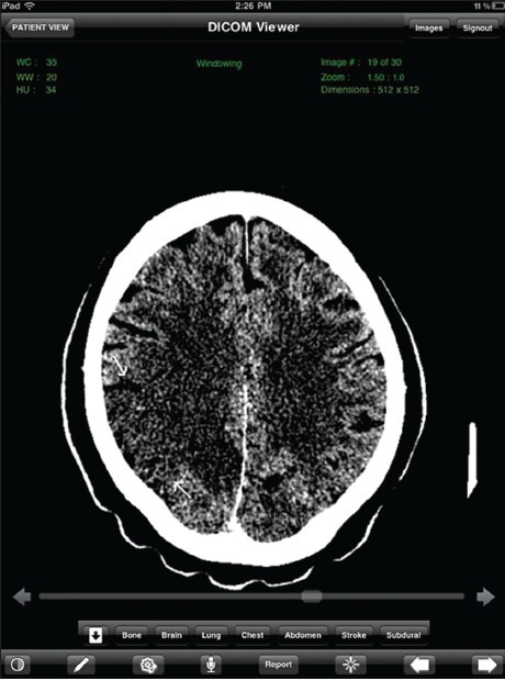
In a study from Massachusetts General Hospital, Gupta et al., evaluated 30 ICU patients who had just had a tube or line placed and concluded that the image quality and accuracy of interpretation is excellent when iPad-based application is used for visualizing chest radiographs and it can potentially aid radiologists for making clinical decisions as there is no delay in downloading images onto the device and time taken to interpret the image is nearly equivalent to the PACS workstation.[11] In another retrospective study on the use of iPad in 20 patients with acute stroke, the same presenters concluded that image quality and accuracy of interpretation is excellent when iPad is used for visualizing stroke images and it overcomes the limitations of iPhone by providing a greater field of view and better download speed, thereby resulting in lesser time for image navigation and evaluation [Figure 1].[12]

A 43-year-old man presented with dizziness and blurred vision. iPad display of noncontrast CT image of the supraventricular brain at narrow window level settings clearly demonstrates an ill-defined hypodensity in the right parietal lobe with loss of gray–white interface (white arrow), consistent with acute infarct
In a study on 120 chest radiographs performed for tuberculosis (TB) screening, Abboud et al., reported no statistically significant difference in sensitivity and specificity for detection of TB between LCD PACS monitors and iPad display.[13]
In a study evaluating 88 on-call CT and magnetic resonance imaging (MRI) studies, John, et al., reported a combined discrepancy rate of 3% for major findings and 5% for minor findings, and concluded that the iPad has the potential to be used safely as an image review tool for on-call CT and MRI studies.[14]
In a study from Italy evaluating 274 lung nodules on chest CT using the iPad and iMac, Faggioni, et al., reported that all lesions detected on the iMac were also found and correctly localized on the iPad2. They further concluded that image reading is relatively fast, supporting the hypothesis that the iPad2 could be reliably used for preliminary visualization of lung nodules.[15]
And finally, in a study from Israel evaluating 134 emergently performed head CTs in a wide spectrum of pathologies including bleed, stroke, and tumor, Shreter et al., reported that all acute findings were correctly interpreted by the reviewing radiologists using the tablet computer.[16]
The message conveyed clearly in the course of the session is that the diagnostic accuracy of radiologists using tablet PCs is in no way inferior to the use of desktop workstations. Any questions that arose were more related to their usability and the efficiency of the reading process using these devices, which currently continue to evolve.
User Feedback
We conducted studies at our institution to assess the efficacy of the iPad in detection of intracranial hemorrhage and pulmonary thromboembolism.[17,18] As part of the study process, the evaluating radiologists were asked to provide feedback on their user experience [Figures [Figures22 and and33].

A 70-year-old male with history of syncope. iPad display of noncontrast CT image at the level of the frontal horn of the lateral ventricle at subdural window settings clearly shows a left frontal convexity acute subdural hematoma (white arrow)

A 58-year-old male presented with chest pain. iPad display of an image of a CT angiogram of the chest demonstrates a filling defect (white arrow) in the left lower lobe pulmonary artery, consistent with pulmonary thromboembolism
Some of the constraints expressed were:
- Window level manual setting is currently slightly cumbersome, using the touch screen feature. Preset windows are more effective.
- Given the small screen size, it is difficult to view two series simultaneously or compare the current study with the prior.
- It is somewhat difficult to measure very small lesions, less than 5 mm in size. As a result, the size of a ureteric calculus can be overestimated which could potentially affect the management of the patient.
However, it was also noted that with increasing use of the iPad, there came a corresponding increase in the comfort level and convenience of its use.
Conclusions
With looming radiologist shortages, an increased demand for imaging, and constantly increasing clinical expectations of instant reporting of emergency cases, the need for paradigm changing technologies such as the IPad becomes more acute. The iPad’s arrival on the world stage is therefore a timely phenomenon from a radiology perspective no less than others.
As the recent RSNA demonstrates, research has shown the quality of image analysis on the iPad to be equal to that of desktop workstation in a variety of clinical applications, including chest radiographs for pneumothorax, chest CT for lung nodules, and CT for acute stroke. As the technology continues to advance, in terms of further improvements in display quality and advanced applications to perform 3D processing on tablet devices, further enhancements will become apparent. We feel that it is only a matter of time before we see mainstream usage of these devices by the radiologist as well as the medical community to access, view, share, and report medical images.
Acknowledgements
The authors acknowledge the input of Mr Raghavendra and Mr Ricky Bedi of TeleradTech in writing this article. The invaluable editorial input by Dr IK Indrajit is also gratefully acknowledged.
Link for the publication: https://www.ncbi.nlm.nih.gov/pmc/articles/PMC3698883/